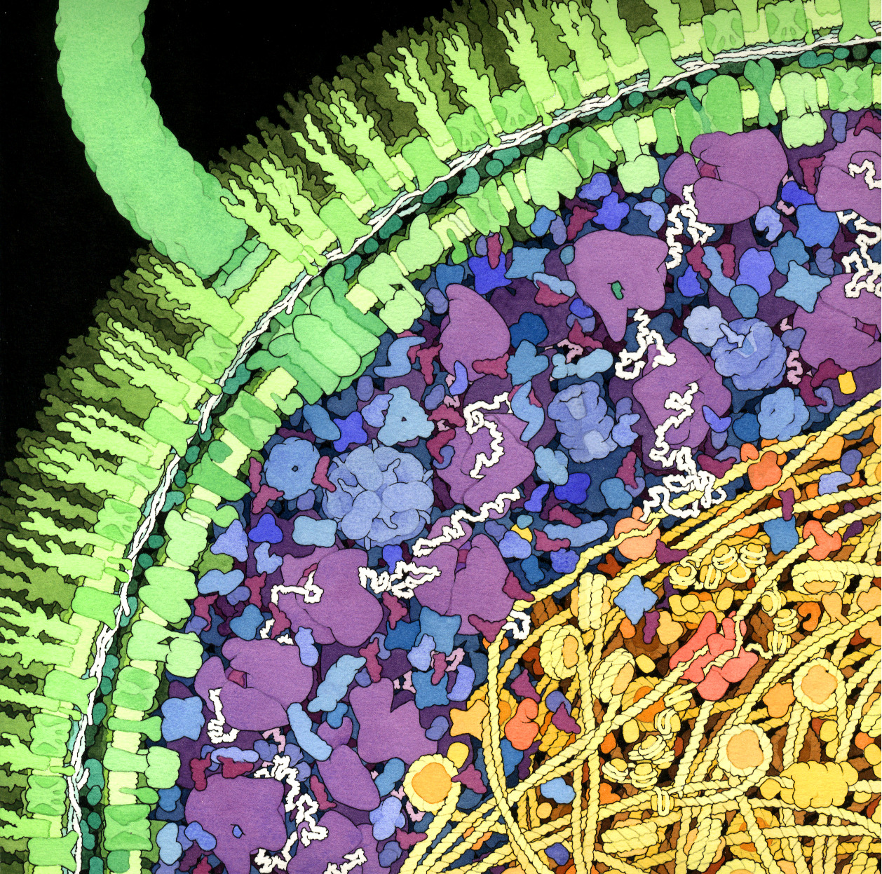
This drawing by scientist and artist David Goodsell shows a cross-section of a small portion of an Escherichia coli cell, giving a hint of the intricate dense disordered complexity of one of the smallest living organisms. Shown in green is the cell wall, with two concentric membranes studded with transmembrane proteins. In the left upper corner, a large flagellar motor crosses the entire wall, turning the flagellum (thick green tube curving up to the vertical) that is many times the length of the bacterium. The cytoplasmic area is colored blue and purple. The large purple molecules are ribosomes (asynchronous parallel synthesis!), the small, L-shaped maroon molecules are tRNA, and the white strands are mRNA. Enzymes are shown in blue. The nucleoid region is shown in yellow and orange, with the long circular DNA shown in yellow, wrapped around HU protein (bacterial nucleosomes). In the center of the nucleoid region, there is a replication fork, in which a red-orange DNA polymerase replicates the new DNA.
This drawing raises many deep and interesting questions of great interest to biophysicists, as well as to biologists, chemists, and even to computer scientists. Some examples are: